ABSTRACT
The ability to manipulate and probe biomolecules at the single-molecule level has become an essential approach for understanding molecular interactions, conformational dynamics, and nanoscale transport phenomena. Advances in experimental techniques have enabled precise control of individual molecules with high spatial resolution and piconewton-level force sensitivity. These developments have significantly expanded the capability of studying biomolecular mechanics and dynamics beyond conventional ensemble measurements. A variety of physical strategies have been developed for single-molecule manipulation, including mechanical-force-based approaches, electric-field-driven methods, and nanoscale structural confinement techniques. Mechanical-force-based methods, such as optical tweezers, magnetic tweezers, and atomic force microscopy, enable direct measurement of molecular mechanical responses. Electric-field-based manipulation, represented by dielectrophoresis, allows noncontact control of particles and biomolecules through polarization effects in non-uniform electric fields. In addition, nanopore-based systems employ nanoscale confinement to regulate molecular transport and residence behavior. This review provides an overview of representative single-molecule manipulation techniques based on mechanical, electrical, and structural control and discusses their fundamental principles and implementation strategies.
-
KEYWORDS: Single-molecule force spectroscopy, Dielectrophoresis, Nanopore trapping, Biomolecular control
1 Introduction
Precise control at the molecular scale has become a fundamental capability in fields ranging from biomolecular analysis to nanoscale characterization [
1,
2]. The ability to manipulate molecules through externally applied physical interactions plays a critical role in determining experimental reproducibility and measurement sensitivity. Over the past decades, a variety of physically driven techniques have been developed to enable controlled interaction with individual molecules and nanoscale analytes [
3–
5].
Physically driven molecular control technologies can be understood as approaches that regulate molecular position, motion, or mechanical state through externally applied forces, electromagnetic fields, or geometrical constraints. Such control extends beyond simple immobilization and includes molecular stretching, confinement, guided transport, and force-mediated modulation. These platforms provide an experimental foundation for quantitative interrogation of molecular behavior [
3].
Existing physically driven molecular control techniques may be structurally organized according to their dominant control strategy. While the detailed operating principles of individual techniques differ, focusing on how molecular control is implemented allows the field to be broadly categorized into three major strategies: (i) externally applied force-based control, (ii) electric-field-induced control, and (iii) structure-induced confinement-based control [
3–
5].
In this review, we systematically examine these physically driven molecular control strategies, focusing on their operating principles, implementation characteristics, and representative applications. By organizing the field from a strategy-oriented perspective, this review aims to provide a structured framework for understanding and selecting appropriate molecular control platforms for diverse experimental objectives.
2 Force-Based Molecular Control Strategies
Force-based molecular control strategies rely on the application of external mechanical forces to individual molecules and the quantitative analysis of their responses. This approach has been primarily developed through single-molecule force spectroscopy (SMFS), which includes representative platforms such as optical tweezers (OT), magnetic tweezers (MT), and atomic force microscopy (AFM) [
6]. Although these techniques differ in their physical implementations, they share a common strategy of regulating molecular position and mechanical state through externally applied forces.
A defining feature of SMFS is its ability to apply piconewton-scale forces while simultaneously measuring nanometer-scale displacements with high precision [
6]. This capability enables detailed investigation of molecular elasticity, conformational transitions, and intermolecular interactions. Consequently, SMFS functions not only as a trapping technique but also as a platform for controlled manipulation of individual biomolecules under mechanical loading. Such measurements have been widely applied to studies of protein unfolding, DNA stretching, and the force-dependent behavior of receptor–ligand interactions [
6].
OT uses optical gradient forces generated by a tightly focused laser beam to trap dielectric beads and apply non-contact forces to tethered molecules [
7,
8]. MT applies forces and torques to magnetic beads through magnetic field gradients, enabling stable control of molecular extension and torsion [
9,
10]. AFM relies on the mechanical interaction between a cantilever tip and a surface-attached molecule, where cantilever deflection provides precise force–distance measurements [
11,
12].
Despite their different operating principles, OT, MT, and AFM share a common framework in which externally applied mechanical forces are used to regulate molecular states and probe their responses. This force-mediated control strategy is fundamentally distinct from other molecular control approaches that rely on electric-field-driven transport or geometrical confinement.
2.1 Optical Tweezers
OT is one of the most widely used force-based molecular control platforms in SMFS. Since the first demonstration of optical trapping of dielectric particles using an infrared laser by Ashkin and co-workers in the mid-1980s [
6], OT has evolved into a powerful tool for applying controlled mechanical forces and analyzing molecular responses at the single-molecule level. In typical experiments, biomolecules are tethered to micrometer-sized optically trapped beads, allowing precise non-contact force application in aqueous environments. This capability makes OT particularly suitable for investigating force-dependent biological processes under conditions close to physiological environments.
OT operates through optical forces generated by momentum transfer between photons and dielectric particles, enabling stable trapping and manipulation of the bead. Unlike contact-based techniques, this mechanism allows relatively smooth and continuous force application. OT typically operates in the force range of approximately 0.1–100 pN while maintaining nanometer-scale spatial resolution and sub-millisecond temporal resolution [
7,
8]. These characteristics position OT as a stable molecular control platform within the intermediate force regime among SMFS techniques.
2.1.1 Fundamental Principles
The physical basis of OT lies in the interaction between a tightly focused laser beam and dielectric particles whose refractive index is higher than that of the surrounding medium. When such particles are placed in a non-uniform optical field, they experience forces arising from the momentum transfer of photons. These forces are commonly decomposed into gradient forces and scattering forces [
7,
8,
13]. The gradient force pulls the particle toward the region of highest optical intensity near the focal point, whereas the scattering force pushes the particle along the direction of beam propagation due to radiation pressure.
Stable three-dimensional trapping is achieved when the gradient force dominates over the scattering force, resulting in the formation of an effective optical potential well [
7,
13]. Near the equilibrium position of the trapped particle, this potential can be approximated as a harmonic potential, in which the restoring force is proportional to the displacement of the particle from the trap center. This harmonic approximation provides the conceptual framework for describing the mechanical behavior of trapped particles [
7,
8].
Within this framework, the proportionality between restoring force and displacement is defined by the trap stiffness, which determines how strongly the particle is confined within the optical trap. The concept of trap stiffness therefore plays a central role in relating optical confinement to mechanical response, enabling controlled manipulation of microscopic objects under optical forces [
7,
8].
In single-molecule experiments, biomolecules are typically tethered between a surface and an optically trapped bead. By controlling the trap position or adjusting the laser intensity, mechanical forces can be applied to the molecule in a well-defined manner. This ability to translate optical forces into controlled mechanical loading forms the fundamental principle underlying OT-based molecular control [
8,
13].
2.1.2 Implementation and Calibration
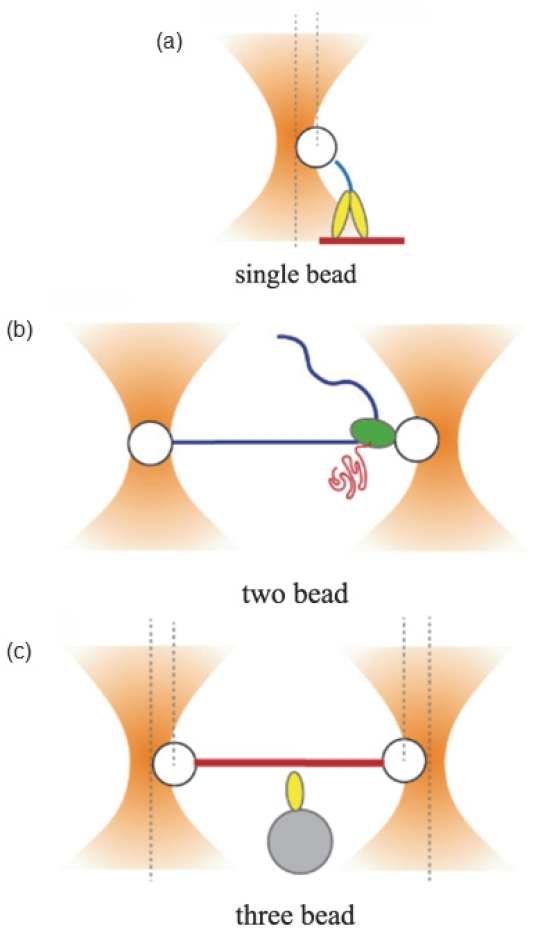
In OT experiments, biomolecules are typically attached to optically trapped microspheres to apply controlled mechanical forces. The molecule is commonly tethered either between a bead and a surface or between two beads, and the specific bead configuration can be tailored depending on the experimental objective. Representative bead configurations used in single-molecule OT experiments are illustrated in
Fig. 1.
In practical systems, the optical trap behaves as a harmonic potential only within a limited displacement range near the focal point. The proportionality constant between restoring force and displacement, known as the trap stiffness, determines the sensitivity and precision of force measurements. Therefore, accurate quantification of trap stiffness is essential for reliable single-molecule force measurements. Because trapped beads undergo continuous Brownian motion in fluid environments, calibration procedures rely on the statistical properties of thermal fluctuations. The equipartition method estimates trap stiffness from the relationship between positional variance and thermal energy, whereas power spectrum analysis extracts trap parameters from the frequency-dependent motion of the bead [
7]. In addition, the trap force constant can be determined by applying a known viscous drag force through controlled movement of the sample stage [
8].
The accuracy of these calibration procedures depends strongly on the stability of optical alignment, precise control of laser power, and reproducible surface functionalization of beads for molecular attachment. Variations in refractive index, temperature drift, and local laser-induced heating can alter trap stiffness and introduce systematic measurement errors [
8]. Consequently, careful calibration is a critical requirement for ensuring quantitative reliability in OT-based single-molecule experiments.
2.1.3 Perspective and Limitation
Although OT provides high precision in force-based molecular manipulation, it also has intrinsic physical limitations arising from the optical trapping mechanism. The trap stiffness is determined by the degree of laser focusing and the gradient of the optical field, and it depends strongly on the size and refractive index of the trapped particle [
7,
8]. For nanoscale objects approaching the diffraction limit, the optical gradient force decreases significantly, leading to reduced trapping efficiency. As a result, the operational force range of conventional far-field OT systems is generally limited to the order of several tens of piconewtons [
7]. In addition, trapped beads in fluid environments undergo continuous Brownian motion, which appears as positional noise and limits the precision of force measurements [
7]. Thermal fluctuations are unavoidable physical factors and therefore impose a fundamental limit on fine force control. Consequently, accurate trap calibration and noise analysis are essential procedures for quantitative single-molecule measurements [
7,
8].
Photothermal effects also represent an important consideration. A portion of the laser energy can be absorbed by the bead or surrounding medium, leading to local temperature increases that may affect sensitive biomolecular systems. These effects become more pronounced during long-duration experiments or under high laser power conditions [
8]. Moreover, slight variations in optical alignment or fluctuations in laser intensity can alter trap stiffness and affect the reproducibility of experimental measurements.
Recent studies have attempted to overcome these limitations by enhancing the optical field gradient or improving trapping efficiency at the nanoscale. In particular, plasmonic nano-optical tweezers have demonstrated the ability to generate strongly localized optical fields, enabling the trapping of nanoscale particles beyond the conventional diffraction limit [
9,
14]. These approaches extend the principles of optical trapping and represent efforts to expand the operational range of OT toward smaller length scales.
MT represent one of the widely used force-based molecular manipulation techniques in single-molecule force spectroscopy. Unlike OT, which relies on optical field gradients, MT generates mechanical forces through magnetic field gradients acting on superparamagnetic beads. Because the magnetic field directly acts on the bead, a constant stretching force can be applied to the molecule for extended periods. Furthermore, by controlling the orientation of external magnets, MT enables the application of torque, allowing the twisting state of biomolecules to be precisely controlled [
10,
15].
Since force generation is decoupled from optical trapping, MT experiments are largely free from photothermal effects associated with laser irradiation. This characteristic makes MT particularly suitable for long-term studies of force-dependent structural transitions and torque-mediated dynamics of biomolecules such as nucleic acids and proteins [
10,
16].
2.2.1 Fundamental Principles
MT are force-based molecular manipulation techniques that utilize the magnetization of superparamagnetic beads in external magnetic fields and the directional forces generated by magnetic field gradients [
10,
15,
16]. In a typical configuration, a biomolecule forms a tether between a surface and a magnetic bead, allowing the magnetic force acting on the bead to be transmitted directly to the molecule. Unlike restoring-force-based systems, the magnitude of the applied force in MT is primarily determined by the arrangement of magnets and the resulting magnetic field distribution rather than by bead displacement.
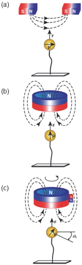
Figure 2 illustrates representative magnetic field configurations used in MT systems. External magnets generate magnetic field gradients that exert stretching forces on the superparamagnetic bead while simultaneously defining the direction of the applied force [
17]. Depending on the magnet geometry, different field orientations can be produced. For example, horizontally arranged magnets induce a horizontal magnetic moment in the bead, whereas cylindrical magnets generate vertical magnetic fields along the tether axis. Magnetic torque tweezers further introduce controlled rotational fields that enable torque-based manipulation of tethered biomolecules [
17].
Because of this mechanism, molecules in MT experiments experience nearly constant tension over time, creating experimental conditions that approximate constant-force environments. In contrast to optical tweezers systems, where harmonic optical potentials generate position-dependent restoring forces, MT defines the force scale primarily through the external magnetic field configuration itself [
10,
15]. In addition to linear stretching forces, MT can also apply rotational control. Rotation of the magnets aligns the magnetic moment of the bead with the magnetic field direction, thereby transmitting torque to the tethered molecule. This capability enables experiments that induce structural transitions such as DNA supercoiling and other twist-dependent conformational changes [
10,
16,
18]. The ability to simultaneously control both force and torque highlights the versatility of MT as a molecular manipulation platform capable of regulating multiple structural degrees of freedom [
10,
16].
2.2.2 Implementation and Calibration
In MT experiments, the magnitude of the applied force is primarily determined by the distance between the magnets and the sample, the magnet arrangement, and the resulting magnetic field gradient [
10,
15]. This configuration allows relatively constant tension to be maintained on the molecule over extended periods, which is particularly advantageous for tracking long-term single-molecule dynamics [
10,
15,
16].
Force calibration is typically performed by analyzing the thermal fluctuations of the superparamagnetic bead [
10,
15]. Under a constant tension, the transverse positional variance of the bead is directly related to the applied force on the molecule, enabling quantitative estimation of the molecular tension [
10,
15]. This approach differs from optical tweezers calibration, where trap stiffness is defined through a harmonic potential, whereas in MT the magnetic field configuration and tether mechanics play central roles in force quantification [
10].
Magnetic bead–based manipulation strategies have also been implemented in microfluidic environments for biomolecular capture and separation. In such systems, externally applied magnetic fields are used to control functionalized magnetic beads within microchannels, enabling selective capture and manipulation of biomolecules without the need for external flow systems [
19].
Recent developments have further extended MT experiments beyond single-tether measurements to highly parallel configurations in which multiple tethers can be simultaneously monitored within a single field of view [
20]. Such parallel measurements improve statistical reliability while emphasizing the importance of tether uniformity and accurate bead tracking for achieving high experimental precision [
10,
20].
2.2.3 Perspective and Limitation
Although MT provides advantages in long-term force stability and torque control, the maximum force that can be applied in typical configurations is generally lower than that achievable with AFM-based methods [
10,
15]. In addition, MT measurements rely on optical tracking of bead motion, and therefore the temporal resolution is limited by the frame rate of the camera and the performance of the optical detection system [
10,
15].
The absolute magnitude and uniformity of the applied force can be affected by several factors, including the magnetic field distribution, variations in the magnetic properties of beads, and the geometric conditions of the tether [
10,
15]. In highly parallel MT configurations where multiple tethers are analyzed simultaneously, these factors may introduce experimental variability, making accurate tether identification and data filtering essential for reliable analysis [
10,
20]. Furthermore, MT experiments typically rely on surface-anchored tether geometries, which may introduce artifacts arising from nonspecific surface interactions or near-surface effects [
10,
15]. Therefore, careful surface passivation and reproducible tether formation are critical for maintaining the quantitative reliability of single-molecule measurements [
10].
AFM is one of the most widely used force-based platforms in SMFS and is capable of probing a broad range of forces. In AFM-based SMFS, a nanoscale probe located at the end of a cantilever forms a mechanical linkage with a single molecule immobilized on a substrate. As the probe retracts, the molecule is stretched and the resulting forces associated with molecular extension or bond rupture are quantitatively measured [
11,
12,
21]. Because AFM relies on a contact-based measurement scheme, it can access relatively high force regimes ranging from tens of piconewtons to several nanonewtons. This capability has enabled extensive studies of protein unfolding, receptor–ligand dissociation, and the mechanical stability of molecular complexes [
12,
21–
23]. In particular, the force–extension curves obtained from cantilever deflection measurements provide direct insights into structural transitions and mechanical responses of individual biomolecules [
22,
23]. Due to these characteristics, AFM occupies the high-force regime among SMFS techniques, complementing the lower-force ranges typically explored by OT and MT [
11,
12,
22].
2.3.1 Fundamental Principles
The fundamental principle of AFM-based SMFS relies on force detection through elastic deformation of the cantilever. The cantilever is typically modeled as an elastic spring, and the force exerted on the molecule is determined by measuring the cantilever deflection when the probe interacts with the molecule [
21,
22]. According to Hooke’s law, the relationship between force and displacement is expressed as
where k represents the spring constant of the cantilever and Δx is the cantilever deflection [
21,
22].
During the experiment, as the probe retracts from the surface, the molecule is progressively stretched. When a critical force is reached, events such as protein domain unfolding or molecular bond rupture occur. These events appear as characteristic signatures in the force–extension curves, enabling quantitative analysis of molecular mechanical stability, folding structures, and interaction energies [
22–
24].
AFM has therefore become a powerful tool for investigating not only the mechanical properties of individual biomolecules but also force-dependent molecular interactions such as receptor–ligand binding and protein folding dynamics [
12,
23].
2.3.2 Implementation and Experimental Configuration
In AFM-SMFS experiments, a stable tether configuration is required to connect a single molecule between the cantilever probe and the substrate surface. Typically, the molecule is immobilized on the substrate, and a mechanical linkage is formed when the AFM probe interacts with the molecule. As the probe retracts from the surface, the molecule is gradually stretched, and the resulting force is measured through cantilever deflection [
12,
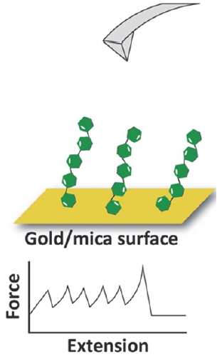
23]. This configuration enables analysis of molecular-scale mechanical events through force–extension curves while maintaining the molecule in a single tether geometry between the probe and the substrate. The general experimental configuration and the force measurement process are illustrated in
Fig. 3. The cantilever tip engages with a surface-anchored molecule and applies a controlled mechanical pulling force as the probe moves away from the substrate, while the deflection of the cantilever provides a quantitative measure of the applied force. Depending on the pulling velocity or force application mode, the unfolding pathway and bond dissociation dynamics of the molecule may vary, providing insights into the energy landscape and mechanical stability of biomolecular systems [
22–
24].
2.3.3 Perspective and Limitation
Although AFM-based SMFS provides access to high-force regimes and high spatial resolution, several limitations arise from its contact-based measurement scheme. Because a direct mechanical contact between the probe and the molecule is required, nonspecific interactions or surface effects may influence the experimental results [
12,
22].
The accuracy of force measurements also strongly depends on proper calibration of the cantilever spring constant and the reproducibility of probe–molecule attachment. Variations in cantilever mechanical properties or surface chemistry can introduce uncertainties and increase experimental variability [
21,
23].
Furthermore, AFM measurements typically analyze one molecular event at a time, which limits experimental throughput. As a result, statistically meaningful results require repeated measurements, and careful data filtering and event classification are essential steps in the analysis of AFM-SMFS experiments [
22–
24].
3 Field-Based Molecular Control Strategies
While force-based molecular control strategies rely on the direct application of mechanical forces to induce molecular extension or structural changes, field-based molecular control approaches manipulate the motion and positioning of particles or biomolecules through polarization effects generated in non-uniform electric field. These strategies enable non-contact manipulation of molecular behavior through externally applied electric field. Among these approaches, dielectrophoresis (DEP) is the most widely studied technique, in which the motion of particles or biomolecules is induced by polarization forces generated in spatially non-uniform electric field.
3.1 Dielectrophoresis (DEP)
DEP is an electric-field-mediated molecular control technique based on the motion of particles induced by polarization forces in non-uniform electric field. DEP can act on electrically neutral particles, where the force arises from polarization induced by differences in dielectric properties between the particle and the surrounding medium [
25,
26]. This characteristic enables spatial manipulation of micro- and nanoscale objects without mechanical contact.
3.1.1 Fundamental Principles
The fundamental principle of DEP lies in the polarization of particles induced by an external electric field. In a uniform electric field, particles do not experience net translational motion; however, when the electric field is spatially non-uniform, polarized particles move along the gradient of electric field energy [
25]. Particles moving toward regions of higher electric field intensity are said to experience positive DEP, whereas those moving toward lower field regions exhibit negative DEP behavior [
27–
29]. The magnitude and direction of the DEP force depend on the effective polarizability of the particle relative to the surrounding medium, and this response may vary with the frequency of the applied electric field [
25]. Consequently, DEP should be understood not simply as the application of an electric field, but as a control strategy that involves the combined design of electric field gradients and frequency-dependent dielectric responses.
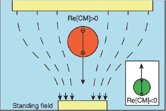
Figure 4 illustrates the basic mechanism of dielectrophoretic motion in a non-uniform electric field. When a particle is placed between electrodes that generate a spatially varying electric field, the electric field induces polarization within the particle, leading to the formation of an induced dipole moment. The interaction between this induced dipole and the electric field gradient produces a net force that drives the particle toward either stronger or weaker electric field regions depending on the relative dielectric properties of the particle and the surrounding medium [
30].
In nanoscale systems, the spatial concentration of electric field gradients becomes a critical factor determining trapping efficiency. Micro- and nanoscale electrode structures, such as graphene-edge electrodes or nanogap architectures, can significantly enhance local electric field gradients and thereby improve the trapping capability for biomolecules [
25–
27].
3.1.2 Implementation and Field Engineering
The implementation of DEP primarily relies on the design of electrode structures that generate non-uniform electric fields. Various configurations, including planar electrodes, microelectrode arrays, and insulator-based DEP devices, have been developed, where electrode geometry and spacing determine the spatial distribution of electric field gradients [
25,
29].
When integrated with microfluidic channels, DEP systems enable simultaneous control of fluid flow and electric field distribution, allowing precise positioning and selective trapping of particles [
31]. Such systems have been applied not only to cells but also to nanoscale particles and biomolecules for controlled assembly and manipulation [
26–
28]. Recent studies have also explored the integration of DEP with sensing platforms by utilizing electric field concentration effects. In these systems, DEP-assisted accumulation of analytes enhances detection sensitivity, extending the role of DEP beyond trapping toward electric-field-assisted sensing mechanisms [
28].
3.1.3 Perspective and Limitation
Although DEP provides a non-contact approach for manipulating particles and biomolecules, its effectiveness strongly depends on the magnitude of electric field gradients and the electrical properties of the surrounding medium [
25]. In high-conductivity environments, electrode heating and electric double-layer effects may occur, potentially affecting structural stability and control precision.
At the nanoscale, polarization forces decrease significantly, making stable trapping difficult without sufficiently strong electric field concentration [
26,
27]. Additional factors such as electrode contamination, electrochemical reactions, and hydrodynamic disturbances may also influence experimental reproducibility [
25,
29]. Therefore, DEP should not be considered merely as the application of an electric field, but rather as a control platform that requires integrated optimization of electrode design, electric field frequency, and medium properties.
4 Structure-Confinement-Based Molecular Control Strategies
While force-based and electric-field-based molecular control strategies rely on externally applied physical fields to manipulate molecular behavior, structure-confinement-based strategies regulate molecular motion through nanoscale geometric constraints. In these approaches, spatial confinement and the resulting free-energy landscape play a central role in controlling molecular dynamics rather than the magnitude of externally applied forces.
Structural confinement can alter molecular trajectories, residence times, and capture probabilities, thereby reshaping the dynamic behavior of molecules. Such approaches enable control over molecular transport without directly applying mechanical forces. Among the platforms in this category, nanopore systems represent one of the most widely studied approaches for structure-based molecular control.
4.1 Nanopore Systems
Nanopores are nanoscale apertures that regulate molecular transport and enable precise control of molecular motion and residence at the single-molecule level. In typical nanopore systems, an electric potential applied across a membrane generates an electric field that drives charged biomolecules toward the nanopore [
32].
Nanopore platforms can be broadly classified into biological nanopores and solid-state nanopores, which differ in material composition, fabrication strategy, and operational characteristics. Biological nanopores, such as α-hemolysin and MspA, are protein-based channels embedded in lipid membranes and offer atomically defined pore structures with high uniformity, enabling low-noise and high-sensitivity detection [
33–
35]. In contrast, solid-state nanopores are fabricated in synthetic membranes using nanofabrication techniques, providing superior mechanical robustness, chemical stability, and tunable geometries, although they typically exhibit higher electrical noise [
36–
38].
A major application of nanopore technology is DNA sequencing, where individual nucleotides are identified by monitoring changes in ionic current as single-stranded DNA translocates through the nanopore. Each nucleotide produces a characteristic modulation of the ionic current, enabling sequence-specific signal discrimination. This label-free approach allows real-time, single-molecule analysis of nucleic acids and has enabled advances such as long-read sequencing, direct RNA sequencing, and portable sequencing platforms for in-field diagnostics [
33–
35].
While early nanopore research focused on translocation-based sensing, recent developments have shifted toward structural confinement strategies that enable prolonged molecular residence and repeated interrogation. This transition reflects the evolution of nanopore systems from passive sensing devices to active platforms for controlling molecular motion through nanoscale confinement [
39–
41].
4.1.1 Fundamental Principles of Structural Confinement
At the nanoscale, molecular transport within nanopores is strongly influenced by geometric confinement. As molecules approach and enter the nanopore, spatial restriction reduces configurational entropy, leading to the formation of free-energy barriers that govern diffusion pathways and residence times [
39–
41]. These effects can result in quasi-stable states near the nanopore entrance. Molecular control in such systems arises from modulation of the free-energy landscape rather than the magnitude of externally applied forces. Entropic effects, electrostatic interactions, and confinement geometry collectively determine key transport parameters, including capture probability, dwell time, and escape dynamics. Thus, nanopore-based control can be understood as an energy landscape engineering approach at the nanoscale.
4.1.2 Implementation and Structural Architectures
Structural confinement in nanopore systems is realized through engineered nanoscale architectures that exploit confinement-induced transport behavior. These approaches regulate molecular motion by introducing geometrical features that control residence and transport dynamics, rather than relying solely on externally applied forces.
Representative implementations include nanofilter structures and confined cavities, which extend molecular residence time by modifying transport pathways [
40,
42]. The nanopore electroosmotic trap (NEOtrap) represents another strategy, where electroosmotic flow stabilizes molecules near the nanopore without direct mechanical confinement [
39].
More advanced designs involve dual-nanopore systems and nanobridge-connected architectures, where two closely spaced nanopores create a confined trapping region between them [
43]. These configurations enable repeated interrogation and long-term observation of individual molecules.
Figure 5 illustrates a representative nanobridge-connected dual nanopore structure, where a nanoscale bridge links adjacent nanopores to form a confined trapping region, enabling stable molecular trapping and electrical detection via ionic current measurements [
43].
4.1.3 Perspective and Limitation
Despite their advantages, nanopore-based molecular control systems face several inherent limitations. Molecular transport is highly dependent on the charge state of analytes and the electrical properties of the surrounding electrolyte. High applied voltages can induce rapid translocation, reducing residence time, whereas low voltages may compromise signal resolution [
32].
Additionally, high ionic strength environments can lead to Joule heating and electric double-layer effects, which may affect nanopore stability and measurement accuracy. Variations in nanopore geometry arising from fabrication processes can also impact reproducibility and trapping efficiency [
5,
32].
Future improvements will require integrated optimization of nanopore geometry, electric field conditions, and fluidic environments. Coupling structural design with electrokinetic control is expected to further enhance performance, enabling more stable and reproducible single-molecule manipulation.
5 Conclusion
In this review, representative strategies for molecular manipulation based on physical control mechanisms were systematically organized. Force-based techniques, including OT, MT, and AFM, enable direct application and measurement of mechanical forces at the single-molecule level. In contrast, DEP manipulates molecular motion through electric-field-induced polarization, while nanopore systems regulate molecular behavior through nanoscale structural confinement. By categorizing these techniques according to their underlying control principles, this review provides a structured perspective for understanding different molecular control platforms and their roles in single-molecule studies.
Notes
-
Acknowledgement
This work was supported by a 2-Year Research Grant of Pusan National University.
-
Conflict of Interest
The authors have no conflicts of interest to declare.
-
Author Contributions
Jeong Hun Shin: Investigation, Writing - Original Draft, Writing.
Tae Won Nam: Review & Editing, Supervision.
Data Availability
Data sharing not applicable – no new data generated.
Fig. 1.Representative optical tweezer bead configurations used in single-molecule experiments: (a) single-bead, (b) two-bead, and (c) three-bead arrangements (Adapted from Ref. [
13], Fig. 47, under CC-BY 4.0 license)
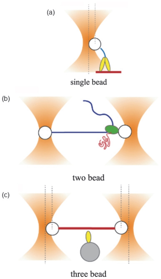
Fig. 2.Representative magnetic field configurations in magnetic tweezers systems. Magnetic field gradients generated by external magnets exert stretching forces on DNA-tethered magnetic beads and enable rotational manipulation depending on the magnet arrangement (Adapted from Ref. [
17] under CC-BY 4.0 license)
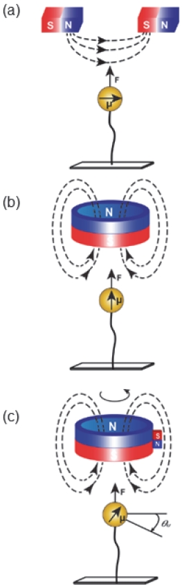
Fig. 3.Representative configuration of AFM-based SMFS. A molecule immobilized on a substrate is mechanically stretched by an AFM cantilever tip, and the resulting force–extension curve reflects molecular unfolding or bond rupture events (Adapted from Ref. [
24], Fig. 1a, under CC BY 4.0 license)
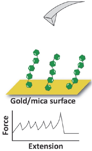
Fig. 4.Dielectrophoretic forces acting on particles in a standing electric field. A particle with positive DEP (red) moves toward regions of higher electric field intensity, whereas a particle with negative DEP (green) is repelled toward regions of lower field intensity (Adapted from Ref. [
30] under CC-BY 4.0 license)
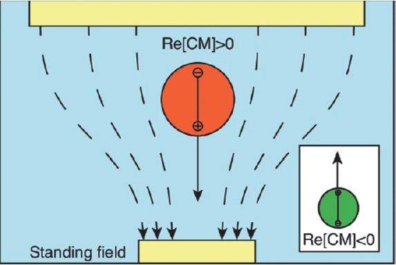
Fig. 5.Schematic representation of a nanobridge-connected dual nanopore configuration formed at the tip of a nanopipette. The nanobridge structure enables confinement of single biomolecules within a nanoscale region between nanopores, allowing stable trapping and electrical detection through ionic current signals (Adapted from Ref. [
43], Fig. 1, under CC BY 4.0 license)
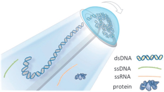
Table 1.Comparison of representative single-molecule control platforms
Table 1.
|
Force range |
Spatial resolution |
Temporal resolution |
Typical applications |
Features |
|
Optical tweezers |
0.1–100 pN |
0.1-2 nm |
10⁴ Hz |
Protein folding, DNA stretching |
Precise force, non-contact |
|
Magnetic tweezers |
0.01–100 pN |
2–10 nm |
10–10² Hz |
DNA twisting, torque studies |
Constant force, parallel |
|
Atomic force microscopy |
10–10⁴ pN |
0.5–1 nm |
10³ Hz |
Protein unfolding, force spectroscopy |
Wide force range, high resolution |
|
Dielectrophoresis |
10–100 pN |
20–800 nm |
10–10³ Hz |
Cell sorting, particle trapping |
Label-free, scalable |
|
Nanopore |
Effective (~pN, field-driven) |
0.1–1 nm |
10³–10⁵ Hz |
DNA sequencing, biosensing |
Electrical readout, real-time analysis |
References
- 1. H. Song, G. Lee, J. Ye, J. Y. Jung, D. Y. Jeong, and J. Ryu, J. Korean Inst. Electr. Electron. Mater. Eng., 37, 119 (2024). doi: https://doi.org/10.4313/JKEM.2024.37.2.1
- 2. D. Park, H. Baek, C. Park, C. Lee, and J. Suh, J. Korean Inst. Electr. Electron. Mater. Eng., 38, 233 (2025). doi: https://doi.org/10.4313/JKEM.2025.38.3.1
- 3. K. C. Neuman and A. Nagy, Nat. Methods, 5, 491 (2008). doi: https://doi.org/10.1038/nmeth.1218
- 4. D. Kim, M. Sonker, and A. Ros, Anal. Chem., 91, 277 (2019). doi: https://doi.org/10.1021/acs.analchem.8b05454
- 5. S. He, Y. Li, S. Fang, Y. Yin, T. Weng, D. Zhou, B. Yin, W. Xie, and D. Wang, Small, 21, e07736 (2025). doi: https://doi.org/10.1002/smll.202507736
- 6. A. Ashkin, J. M. Dziedzic, and T. Yamane, Nature, 330, 769 (1987). doi: https://doi.org/10.1038/330769a0
- 7. K. C. Neuman and S. M. Block, Rev. Sci. Instrum., 75, 2787 (2004). doi: https://doi.org/10.1063/1.1785844
- 8. G. Pesce, P. H. Jones, O. M. Maragò, and G. Volpe, Eur. Phys. J. Plus, 135, 949 (2020). doi: https://doi.org/10.1140/epjp/s13360-020-00843-5
- 9. J. D. Kolbow, N. C. Lindquist, C. T. Ertsgaard, D. Yoo, and S. H. Oh, ChemPhysChem, 22, 1409 (2021). doi: https://doi.org/10.1002/cphc.202100004
- 10. H. K. Choi, H. G. Kim, M. J. Shon, and T. Y. Yoon, Annu. Rev. Biochem., 91, 33 (2022). doi: https://doi.org/10.1146/annurev-biochem-032620-104637
- 11. E. Dague, V. Pons, A. Roland, J. M. Azaïs, S. Arcucci, V. Lachaize, S. Velmont, E. Trevisiol, D. N'Guyen, J. M. Sénard, and C. Galés, Commun. Biol., 5, 221 (2022). doi: https://doi.org/10.1038/s42003-022-03162-w
- 12. S. S. Mandal, ACS Omega, 5, 11271 (2020). doi: https://doi.org/10.1021/acsomega.0c00814
- 13. G. Volpe, O. M. Maragò, H. Rubinsztein-Dunlop, G. Pesce, A. B. Stilgoe, G. Volpe, G. Tkachenko, V. G. Truong, S. N. Chormaic, F. Kalantarifard, P. Elahi, M. Käll, A. Callegari, M. I. Marqués, A. A. R. Neves, W. L. Moreira, A. Fontes, C. L. Cesar, R. Saija, A. Saidi, P. Beck, J. S. Eismann, P. Banzer, T. F. D. Fernandes, F. Pedaci, W. P. Bowen, R. Vaippully, M. Lokesh, B. Roy, G. Thalhammer-Thurner, M. Ritsch-Marte, L. P. García, A. V. Arzola, I. P. Castillo, A. Argun, T. M. Muenker, B. E. Vos, T. Betz, I. Cristiani, P. Minzioni, P. J. Reece, F. Wang, D. McGloin, J. C. Ndukaife, R. Quidant, R. P. Roberts, C. Laplane, T. Volz, R. Gordon, D. Hanstorp, J. T. Marmolejo, G. D. Bruce, K. Dholakia, T. Li, O. Brzobohatý, S. H. Simpson, P. Zemánek, F. Ritort, Y. Roichman, V. Bobkova, R. Wittkowski, C. Denz, G. V. P. Kumar, A. Foti, M. G. Donato, P. G. Gucciardi, L. Gardini, G. Bianchi, A. V. Kashchuk, M. Capitanio, L. Paterson, P. H. Jones, K. Berg-Sørensen, Y. F. Barooji, L. B. Oddershede, P. Pouladian, D. Preece, C. B. Adiels, A. C. De Luca, A. Magazzù, D. Bronte Ciriza, M. A. Iatì, and G. A. Swartzlander, J. Phys. Photonics, 5, 022501 (2023). doi: https://doi.org/10.1088/2515-7647/acb57b
- 14. M. L. Juan, M. Righini, and R. Quidant, Nat. Photon., 5, 349 (2011). doi: https://doi.org/10.1038/nphoton.2011.56
- 15. D. Dulin, Methods Mol. Biol., 2694, 375 (2024). doi: https://doi.org/10.1007/978-1-0716-3377-9_18
- 16. R. Tapia-Rojo, Biophys. Rev., 17, 25 (2025). doi: https://doi.org/10.1007/s12551-025-01274-1
- 17. R. Sarkar and V. V. Rybenkov, Frontiers Phys., 4, 48 (2016). doi: https://doi.org/10.3389/fphy.2016.00048
- 18. V. Croquette, J. Valle-Orero, M. Rieu, and J. F. Allemand, Methods Enzymol., 694, 1 (2024). doi: https://doi.org/10.1016/bs.mie.2024.01.026
- 19. J. Wang, K. Morabito, T. Erkers, and A. Tripathi, Analyst, 138, 6573 (2013). doi: https://doi.org/10.1039/c3an01166f
- 20. I. De Vlaminck, T. Henighan, M. T. J. van Loenhout, I. Pfeiffer, J. Huijts, J. W. J. Kerssemakers, A. J. Katan, A. van Langen-Suurling, E. van der Drift, C. Wyman, and C. Dekker, Nano Lett., 11, 5489 (2011). doi: https://doi.org/10.1021/nl203299e
- 21. F. A. Carvalho and N. C. Santos, IUBMB Life, 64, 465 (2012). doi: https://doi.org/10.1002/iub.1037
- 22. Y. Kim, W. Kim, and J. W. Park, Bull. Korean Chem. Soc., 37, 1895 (2016). doi: https://doi.org/10.1002/bkcs.11022
- 23. M. L. Hughes and L. Dougan, Rep. Prog. Phys., 79, 076601 (2016). doi: https://doi.org/10.1088/0034-4885/79/7/076601
- 24. B. Yang, Z. Liu, H. Liu, and M. A. Nash, Front. Mol. Biosci., 7, 85 (2020). doi: https://doi.org/10.3389/fmolb.2020.00085
- 25. A. Barik, Y. Zhang, R. Grassi, B. P. Nadappuram, J. B. Edel, T. Low, S. J. Koester, and S. H. Oh, Nat. Commun., 8, 1867 (2017). doi: https://doi.org/10.1038/s41467-017-01635-9
- 26. E. S. Yu, H. Lee, S. M. Lee, J. Kim, T. Kim, J. Lee, C. Kim, M. Seo, J. H. Kim, Y. T. Byun, S. C. Park, S. Y. Lee, S. D. Lee, and Y. S. Ryu, Nat. Commun., 11, 2804 (2020). doi: https://doi.org/10.1038/s41467-020-16630-w
- 27. S. Fujiwara, M. Hata, I. Onohara, D. Kawasaki, K. Sueyoshi, H. Hisamoto, M. Suzuki, T. Yasukawa, and T. Endo, RSC Adv., 13, 21118 (2023). doi: https://doi.org/10.1039/d3ra03245k
- 28. Z. Çağlayan, Y. D. Yalçın, and H. Külah, Micromachines, 11, 990 (2020). doi: https://doi.org/10.3390/mi11110990
- 29. M. Oh, V. Jayasooriya, S. O. Woo, D. Nawarathna, and Y. Choi, ACS Appl. Nano Mater., 3, 797 (2020). doi: https://doi.org/10.1021/acsanm.9b02302
- 30. Z. Tian, X. Wang, and J. Chen, Microsyst. Nanoeng., 10, 117 (2024). doi: https://doi.org/10.1038/s41378-024-00750-0
- 31. N. Izquierdo, R. Li, P. R. Christenson, S. H. Oh, and S. J. Koester, ACS Appl. Mater. Interfaces, 17, 32764 (2025). doi: https://doi.org/10.1021/acsami.4c22829
- 32. M. Wanunu, Phys. Life Rev., 9, 125 (2012). doi: https://doi.org/10.1016/j.plrev.2012.05.010
- 33. F. Haque, J. Li, H. C. Wu, X. J. Liang, and P. Guo, Nano Today, 8, 56 (2013). doi: https://doi.org/10.1016/j.nantod.2012.12.008
- 34. Z. L. Hu, M. Z. Huo, Y. L. Ying, and Y. T. Long, Angew. Chem. Int. Ed., 60, 14738 (2021). doi: https://doi.org/10.1002/anie.202013462
- 35. D. Branton, D. W. Deamer, A. Marziali, H. Bayley, S. A. Benner, T. Butler, M. Di Ventra, S. Garaj, A. Hibbs, X. Huang, S. B. Jovanovich, P. S. Krstic, S. Lindsay, X. S. Ling, C. H. Mastrangelo, A. Meller, J. S. Oliver, Y. V. Pershin, J. M. Ramsey, R. Riehn, G. V. Soni, V. Tabard-Cossa, M. Wanunu, M. Wiggin, and J. A. Schloss, Nat. Biotechnol., 26, 1146 (2008). doi: https://doi.org/10.1038/nbt.1495
- 36. S. He, Y. Liu, S. Fang, Y. Li, T. Weng, R. Tian, Y. Yin, D. Zhou, B. Yin, Y. Wang, L. Liang, W. Xie, and D. Wang, Coord. Chem. Rev., 510, 215816 (2024). doi: https://doi.org/10.1016/j.ccr.2024.215816
- 37. Y. He, M. Tsutsui, Y. Zhou, and X. S. Miao, NPG Asia Mater., 13, 48 (2021). doi: https://doi.org/10.1038/s41427-021-00313-z
- 38. W. Liu, J. Andersson, J. Järlebark, A. Shaji, J. Sha, and A. Dahlin, Nano Lett., 25, 9654 (2025). doi: https://doi.org/10.1021/acs.nanolett.5c01447
- 39. S. Schmid, P. Stömmer, H. Dietz, and C. Dekker, Nat. Nanotechnol., 16, 1244 (2021). doi: https://doi.org/10.1038/s41565-021-00958-5
- 40. X. Liu, M. M. Skanata, and D. Stein, Nat. Commun., 6, 6222 (2015). doi: https://doi.org/10.1038/ncomms7222
- 41. S. Pud, S. H. Chao, M. Belkin, D. Verschueren, T. Huijben, C. van Engelenburg, C. Dekker, and A. Aksimentiev, Nano Lett., 16, 8021 (2016). doi: https://doi.org/10.1021/acs.nanolett.6b04642
- 42. M. H. Lam, K. Briggs, K. Kastritis, M. Magill, G. R. Madejski, J. L. McGrath, H. W. de Haan, and V. Tabard-Cossa, ACS Appl. Nano Mater., 2, 4773 (2019). doi: https://doi.org/10.1021/acsanm.9b00606
- 43. P. Cadinu, B. P. Nadappuram, D. J. Lee, J. Y. Y. Sze, G. Campolo, Y. Zhang, A. Shevchuk, S. Ladame, T. Albrecht, Y. Korchev, A. P. Ivanov, and J. B. Edel, Nano Lett., 17, 6376 (2017). doi: https://doi.org/10.1021/acs.nanolett.7b03196